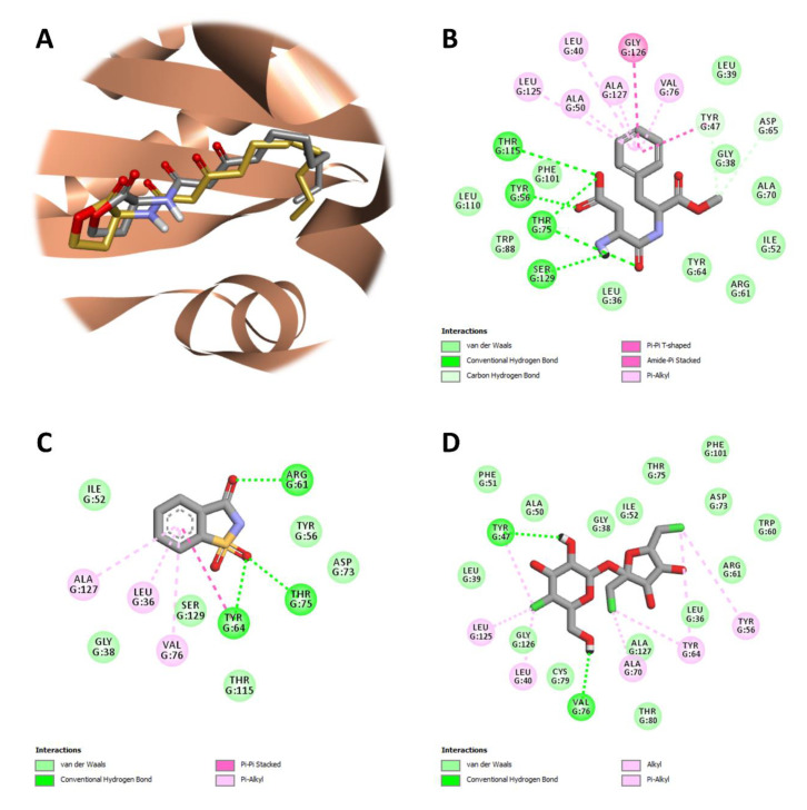
Figure 5.
Protein-ligand docking and interaction profiling. (A) Close-up view of the superposed structures of co-crystallized (gold) and redocked (silver) 3-oxo-C12-HSL in the ligand-binding cavity of LasR (PDB ID: 2UV0; chain ID: G). Favorable non-covalent interactions stabilizing the relative position of (B) aspartame, (C) saccharin, or (D) sucralose with respect to the LasR-LBD. Images were prepared and rendered using Discovery Studio Visualizer, version 16.1.0 (Dassault Systèmes BIOVIA Corp., San Diego, CA, USA).

