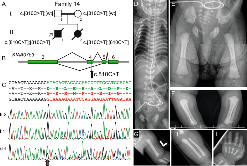
Fig. 2.
Molecular and radiographic features of the proband in family 14 with a homozygous synonymous variant introducing abnormal splicing in KIAA0753 (NM_014804.4). A Pedigree of family 14 and segregation of the KIAA0753 variant. B Schematic figure of the genomic region from exon 3 to 6 in KIAA0753, indicating the normal splice pattern (top) and abnormal splicing (bottom). The black arrow shows the position of the c.810C>T variant in exon 4. C Sequence of exon 3 (text in black), exon 4 (green) and exon 5 (red). cDNA sequence traces show skipping of exon 4 in homozygous state for patient II:2, heterozygous state for patients father, shown by lower peaks of normal isoform transcript in I:1 (sequence from mother I:2 not shown). Analysis of normal control sample shows that the aberrant splicing pattern in very low extent is also present in healthy individuals (n = 3). The red arrow shows where the exon skipping occurs. (D-I) Radiograms of II:2 at 5 days of age. Note narrow thorax, short ribs and characteristic “handle bar” appearance of the clavicles. E–H short craniocaudal diameter of the iliac bones, trident pelvis, short ischial bones and short tubular bones with bulbous ends. The hand shows short metacarpals and phalanges, note that middle phalanges are particularly short and broad (I). ctrl control, wt wildtype

