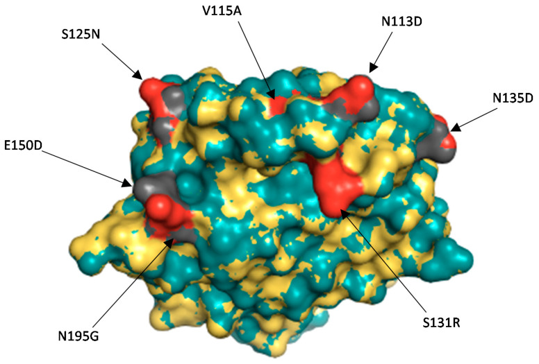
Figure 4.
Surface representation of the VP8* protein of Rotarix® and the divergent study strain UFS-NGS-MRC-DPRU4749. The superposition of the two structures has the root square mean deviation of 0.048 Å. Rotarix® structure is represented by the teal colour whereas the Zambian P[8] strain is indicated in yellow. The red colour represents the amino acid changes observed on the Zambian study strain as compared to Rotarix® vaccine strain in grey.

