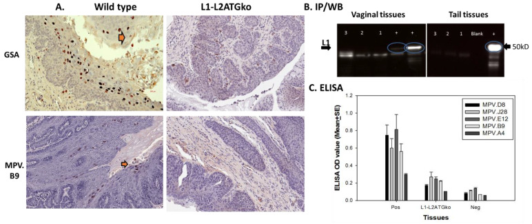
Figure 8.
No L1 protein was detected in L1-L2ATGko infected tissues by IHC, ELISA, and IP/Western. Both wild-type and L1–L2 mutant infected tissues were examined for viral L1 using GSA and MPV.B9 antibody. No positive signals were detected in L1-L2ATGko mutant-induced tissues (A, right panel), while positive signals could be detected in wild-type infected tissues (left panel, arrows). No L1 positive signals were detected in L1-L2ATGko mutant-infected vaginal tissues by direct Western blot (left, lanes 1–3 from three animals, two wild-type lesions as positive controls “+”). No L1 was detected in L1-L2ATGko tail tissues (right, lanes 1–3 from three animals, one wild-type tail lesion as positive control “+”) in an IP/Western immunoprecipitation using MPV.A4 to pull down L1 and probing the blot with MPV.B9 (B). Wild-type and L1-L2ATGko mutant-infected tissues were also tested for L1 by ELISA (C) using a panel of in-house monoclonal antibodies including MPV.B9 and MPV.A4 that were raised against the mouse papillomavirus major capsid protein L1. No significantly difference was found between the L1-L2ATGko mutant and the negative group (p > 0.05, Mann–Whitney rank-sum test).

