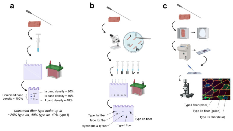
Figure 1.
Summary of techniques. Legend: (a) fiber type estimation based on biopsied homogenates: a biopsy is obtained, the tissue is homogenized in specialized buffers and prepped for electrophoresis, and the gel is stained post-electrophoresis to visualize the percentage of each myosin isoform band. (b) singe fiber analysis: a biopsy is obtained, the tissue is teased apart under a stereoscope in a physiological digestion buffer, every single fiber is placed in a tube and homogenized, and electrophoresis is performed with back-end gel staining; this allows for the confident detection of hybrid fibers (example being “fiber iii”). (c) immunohistochemistry: a biopsy is obtained, the tissue is slow-frozen in a cryomold (or on cork) using freezing media, the frozen tissue is sectioned onto microscope slides using a cryostat, primary antibody solutions against various myosin isoforms are pipetted onto the slide, secondary antibody solutions against the primary antibodies are pipetted onto the slide, and the slide is mounted and imaged on a fluorescent microscope. Note, this image was generated using BioRender.com, and the fluorescent image is from the laboratory of MDR.

