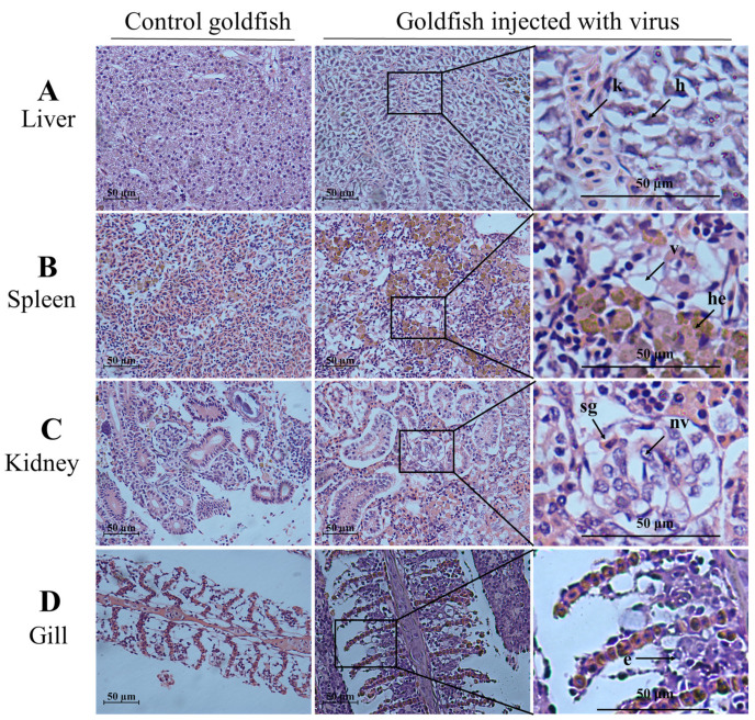
Figure 6.
Histological changes in the liver, spleen, kidney, and gill from challenged goldfish (HE staining). Scale bars = 50 µm. (A) Extensive necrosis of liver parenchyma. Karyopyknosis (k) and hyperemia (h) marked by an arrow; (B) vacuolation of cytoplasm (v) and hemosiderosis (he) in spleen; (C) the arrow shows the swelling of glomerular (sg) and nuclear vacuole (nv); (D) exfoliation of epithelial cells (e) marked by an arrow.

