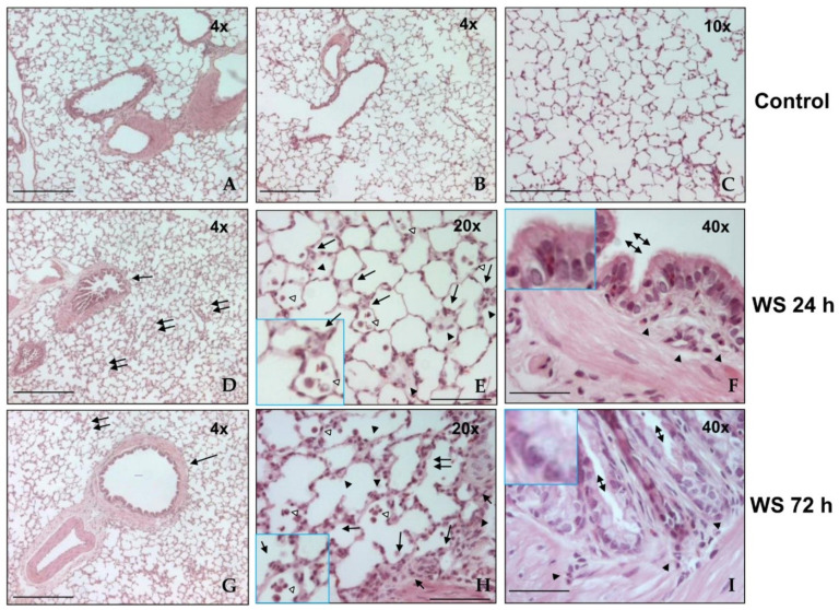
Figure 1.
Representative photomicrographs of histological lung sections from WS-exposed guinea pigs and controls (n = 8). Controls (A–C), after 24 h (D–F), and after 72 h (G–I) of exposure to WS. The bottom-left inserts in panels E and I show intralveolar and alveolar macrophages (empty arrow) and polymorphonuclear leucocytes (arrow). Upper-left inset in panels in F and I display goblet cell hyperplasia. Hematoxylin–eosin stain. Original magnification, panels A, B, D, and G, 4×; panel C, 10×; panels E and H, 2×; panels F and I, 40×. Scale bars = 100 µm.

