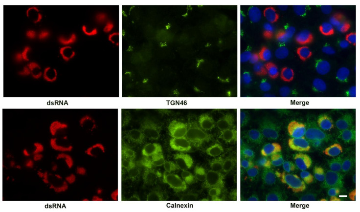
Figure 7.
Immunofluorescent labeling of the SARS-CoV-2 replication complex at 24 h post-infection by antibody detecting dsRNA (red) and co-labeling with antibody to Golgi (TGN46) and ER markers (Calnexin). Cellular antigens are shown in green. The merged image is counterstained with DAPI (blue). Note the condensation of the ER and close association with the viral replication organelle. TGN46 appears only as diffuse background in infected cells. Bar = 10 μm.

