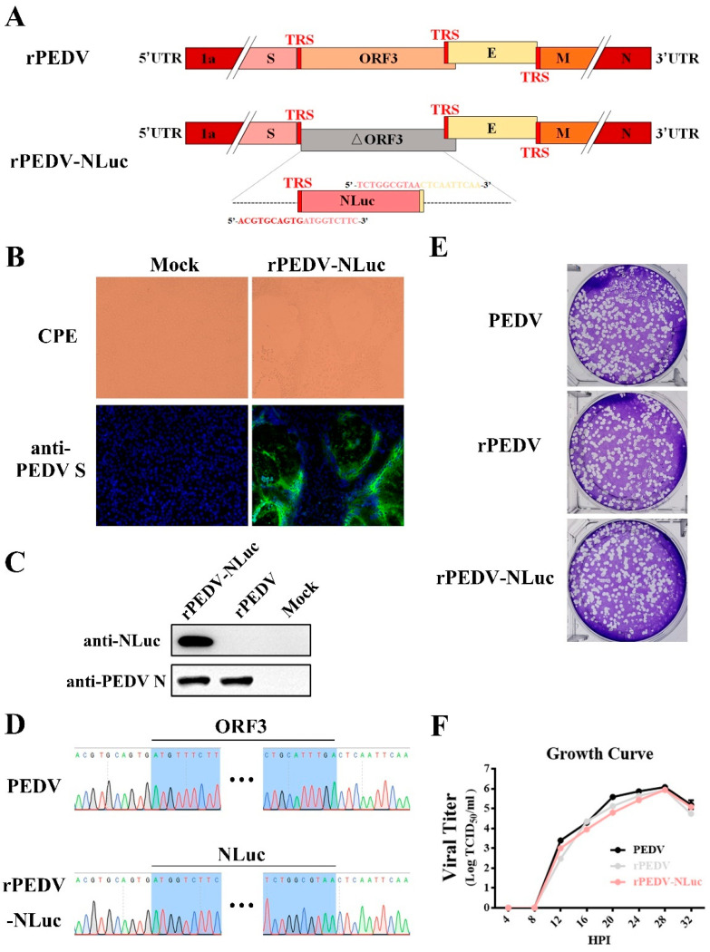
Figure 2.
Rescue of rPEDV-NLuc. (A) Schematic representation of the cDNA clone of rPEDV-NLuc. (B) The rPEDV-NLuc- and mock-infected Vero cells were detected by IFA using anti-PEDV S mAb 4B2 (magnification, ×100). (C) Western blot analysis of the expression of PEDV N and NLuc using the anti-PEDV N mAb 8E2 and the anti-NLuc mAb 5H6, respectively. (D) Identification of the ORF3 replacement with NLuc by sequencing the rPEDV-NLuc genome. (E) Representative plaques of wild-type PEDV-, rPEDV-, and rPEDV-NLuc-infected Vero cell. (F) Comparison of growth kinetics of wild-type PEDV, rPEDV, and rPEDV-NLuc. Vero cells were infected with wild-type PEDV, rPEDV, and rPEDV-NLuc at an MOI of 0.01, and viral titers were detected at indicated time points by TCID50 assay. Data are represented as the mean ± SD (n = 3).

