
FIGURE 3.
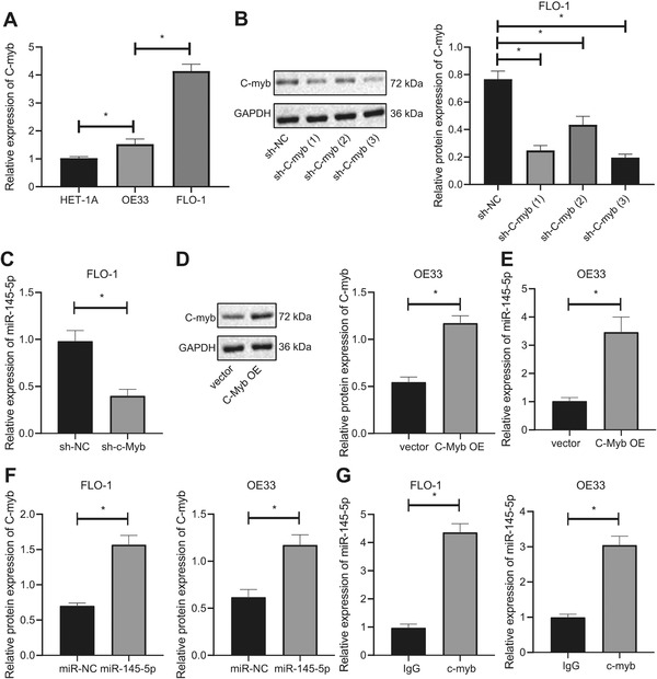
c‐Myb activates the expression of miR‐145‐5p. (A) RT‐qPCR determination of expression of c‐Myb in each EAC cell line and normal esophageal mucosal epithelial cells. (B) Western blot analysis of the expression of c‐Myb in FLO‐1 cells after treatment of sh‐c‐Myb. (C) RT‐qPCR determination of expression of miR‐145‐5p in FLO‐1 cells after treatment of sh‐c‐Myb. (D) Western blot analysis of c‐Myb expression in OE33 cells after treatment of c‐Myb OE. (E) Western blot analysis of expression in OE33 cells after treatment of c‐Myb OE. (F) The binding of miR‐145‐5p to c‐Myb in EAC cells assessed by RNA pull‐down assay. (G) The binding of miR‐145‐5p to c‐Myb in EAC cells through antibodies to IgG and c‐Myb using RIP assay. *P < .05. Measurement data were expressed as the mean ± standard deviation. One‐way ANOVA and Tukey's post‐hoc test were used for data analysis in panels A and B, and unpaired t‐test was employed for data in panels C‐G