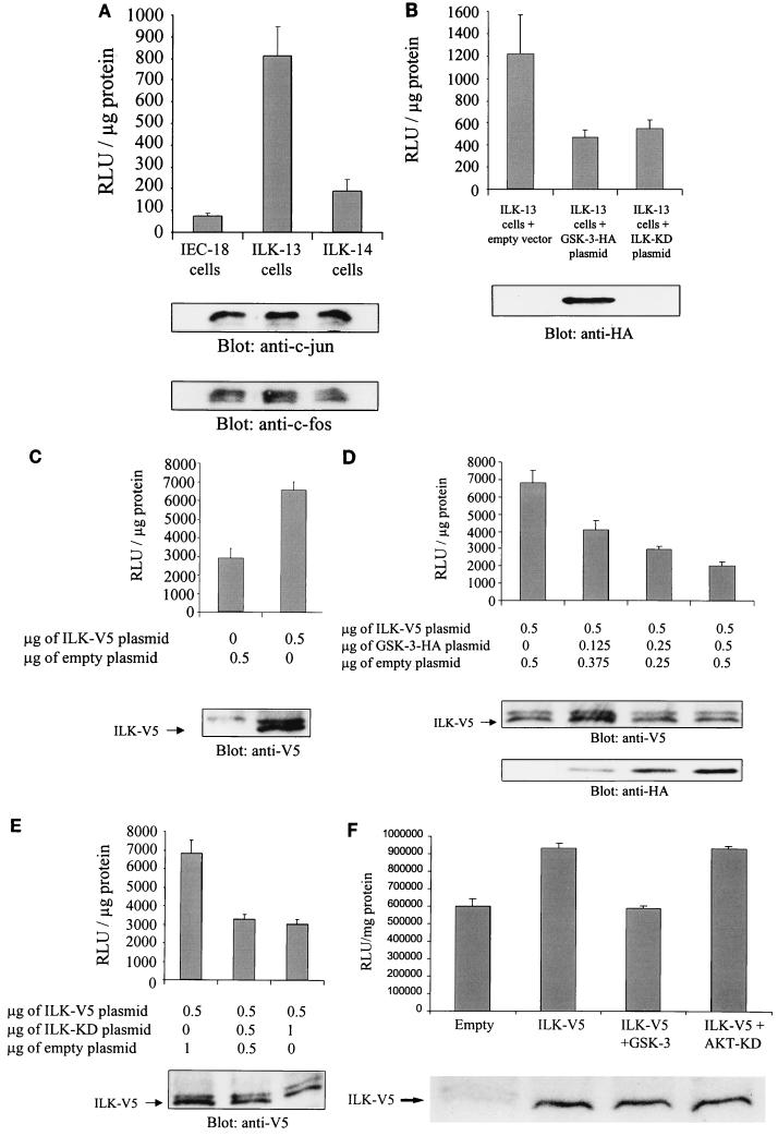
FIG. 2.
ILK regulates AP-1 activity in a GSK-3-dependent manner. (A) AP-1 activity in IEC-18 cells and in IEC-18 cells stably transfected with wild-type ILK (ILK-13; clone A1a3 (37, 40) and anti-sense ILK (ILK-14; clone A2C3) was measured. After transfection of 0.5 μg of pGL3-AP-1 by Lipofectin, AP-1 activity was determined by luciferase assay. c-jun and c-fos protein expression levels in each cell line were estimated by Western blotting. RLu, relative light units. (B) Overexpression of GSK-3 decreases AP-1 activity in ILK-13 cells. Luciferase assays were performed with ILK-13 cells transfected with 0.5 μg of pGL3-AP-1 and 0.5 μg of pcDNA-GSK-3-HA or 0.5 μg of pcDNA-ILK-KD. Transfection and expression of GSK-3 were established by Western blot analysis using an anti-HA antibody. (C) AP-1 activity in HEK-293 cells transiently transfected with ILK. HEK-293 cells were transiently cotransfected with 0.5 μg of pGL3-AP-1 and 0.5 μg of pcDNA3.1-ILK-V5 or empty vector. ILK expression in the transfected cells was detected by immunoblotting with an anti-V5 antibody. (D) Dose-dependent effect of GSK-3 on ILK-induced AP-1 activity. Increasing amounts of pcDNA-GSK-3-HA (0.125, 0.25, and 0.5 μg) were cotransfected with 0.5 μg of pGL3-AP-1 and 0.5 μg of pcDNA3.1-ILK-V5, and AP-1 activity was evaluated. Expression of ILK and GSK-3 in the transfected cells is shown by Western blotting with anti-V5 and anti-HA antibodies, respectively. (E) Effect of ILK-KD on ILK-induced AP-1 activity. The same experiment as that shown in panel D was performed with 0.5 μg and 1 μg of pcDNA-ILK-KD. Expression of ILK in the transfected cells was established by Western blot analysis with an anti-V5 antibody. (F) Effect of AKT-KD on ILK-induced AP-1 activity. HEK-293 cells were cotransfected with 0.5 μg pGL3-AP-1, 0.5 μg of pcDNA3.1-ILK-V5, and 0.5 μg of pcDNA-GSK-3-HA or 0.5 μg of pcDNA-AKT-KD-HA. Expression of ILK in the transfected cells was established by Western blot analysis with an anti-V5 antibody.

