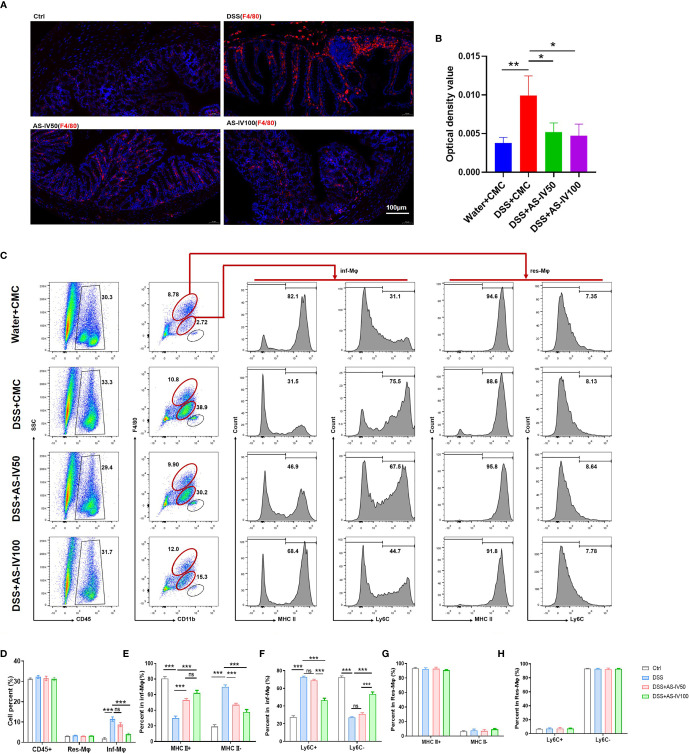
Figure 3.
AS-IV remodeled the development of intestinal macrophages. (A) F4/80 protein expression in colon tissue was detected by immunofluorescence. Positive immunoreactivity for F4/80 protein expression is indicated by the red color. The slides were counterstained with DAPI (blue). In addition, the sum of OD was analyzed in (B). (C) Intestinal macrophages were collected and analyzed by fluorescence-activated cell sorting (FACS) after staining with anti-F4/80, anti-CD11b, and anti-MHCII (or anti-Ly6C). (D–H) The number of the different subpopulations in (C) was determined and quantitatively compared. One-way ANOVA test was used for statistical analyses. Bars represent means ± SD; * P < 0.05, **P < 0.01, *** P < 0.001. ns, no significance.

