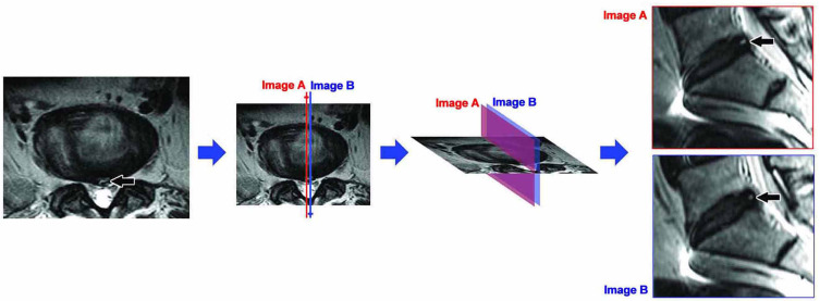
Figure 2.
Consecutive slides HIZ. Axial T2-weighted MR image of the lumbar spine shows a HIZ in the posterior annulus fibrosus at L5S1 (black arrow). Two adjacent sagittal images, slide A (red) and B (blue), show HIZs in similar position at L5S1 (black arrow), ie Consecutive slides HIZ. It indicates the same HIZ lesion on Axial T2-weighted MR image.

