Figure 3.

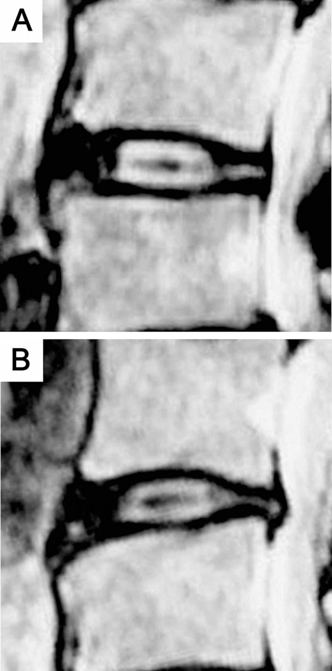
Abnormal signals extending from nucleus pulposus forward or posterior into the signal of the annulus fibrous on sagittal T2-weighted MR images. (A) “Rat tail” abnormal signal; (B) “Mallet head” abnormal signal.

Abnormal signals extending from nucleus pulposus forward or posterior into the signal of the annulus fibrous on sagittal T2-weighted MR images. (A) “Rat tail” abnormal signal; (B) “Mallet head” abnormal signal.