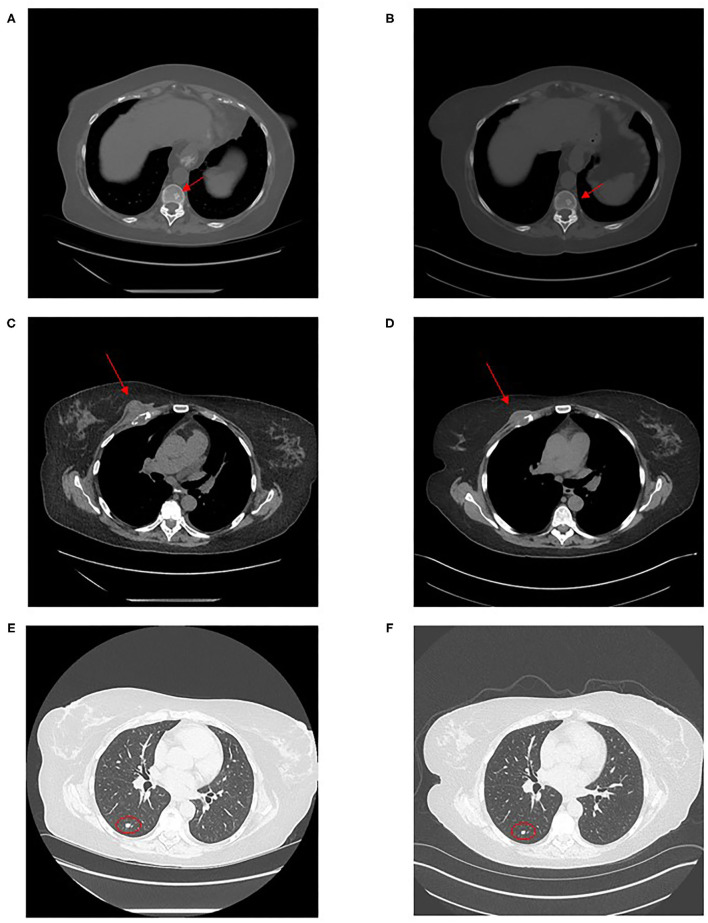
Figure 2.
Computed tomography images of disseminated coccidioidomycosis lesions. Computed tomography images show: an axial sclerotic bone lesion in December 2020 (A) and after 4 months of fluconazole treatment in April 2021 (B), the chest wall mass in December 2020 (C) and in April 2021 (D) and a right lower lobe lung nodule in December 2020 (E) and in April 2021 (F).

