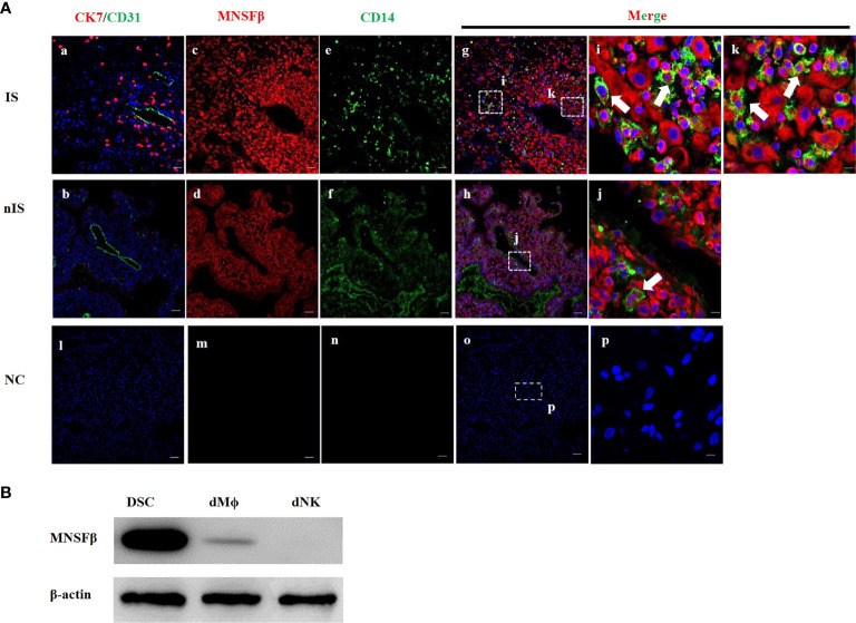
Figure 1.
Expression of MNSFβ at the maternal–fetal interface. (A) Distribution of MNSFβ expression in human decidual tissues of a physically normal pregnant woman in the first trimester (8W) as determined by immunofluorescent staining analysis. (a, b) Staining of CK7 (red, marker of trophoblast cells) and CD31 (green, marker of endothelial cells) to confirm the embryo implantation site (IS) and nonimplantation site (nIS), and the cell nucleus was stained blue. (c, d) Staining of MNSFβ (red). (e, f) Staining of CD14 (green, marker of macrophages). (g, h) Staining of MNSFβ (red) and CD14 (green), and the nucleus was stained blue. (i, k) Magnification of images in panel (g), (j) Magnification of images in panel (h). (l–p) NC (negative control) of CK7/CD31, MNSFβ, and CD14. White arrow indicates typical positive cells. Scale bars of a–h represent 100 μm; scale bars of i–k represent 10 μm. (B) Detection of the MNSFb protein expression in human decidual stromal cells (DSCs), decidual macrophages (dMФ), and decidual NK cells (dNKs) of early pregnancy by Western blot analysis.

