Figure 3.
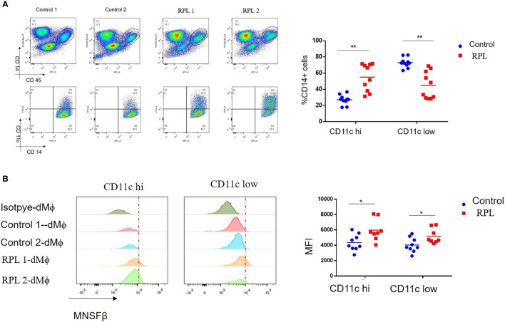
Changes in the proportion of the CD11c high and CD11c low subsets in human dMϕ from RPL patients. (A) Proportion of the CD11c high (CD11c hi) and CD11c low (CD11c low) subsets in the dMϕ of Control women (n = 9) and RPL patients (n = 8) as detected by flow cytometry. Left panel: flow cytometry analysis of dMϕ with antibodies against CD45 plus CD14 and CD14 plus CD11c. Right panel: Proportions of CD11c hi and CD11c low dMϕ. (B) MNSFβ protein expression levels in CD11c hi and CD11c low dMϕ from Control women (n = 9) and RPL patients (n = 8) as detected by flow cytometry. Left panel: Representative images of the flow cytometry assay; right panel: the mean fluorescence intensity (MFI) of MNSFβ as detected by flow cytometry. (Control: dMϕ isolated from decidual tissues of normal women in early pregnancy; RPL: dMϕ isolated from decidual tissues of RPL patients, *p < 0.05, **p < 0.01).

