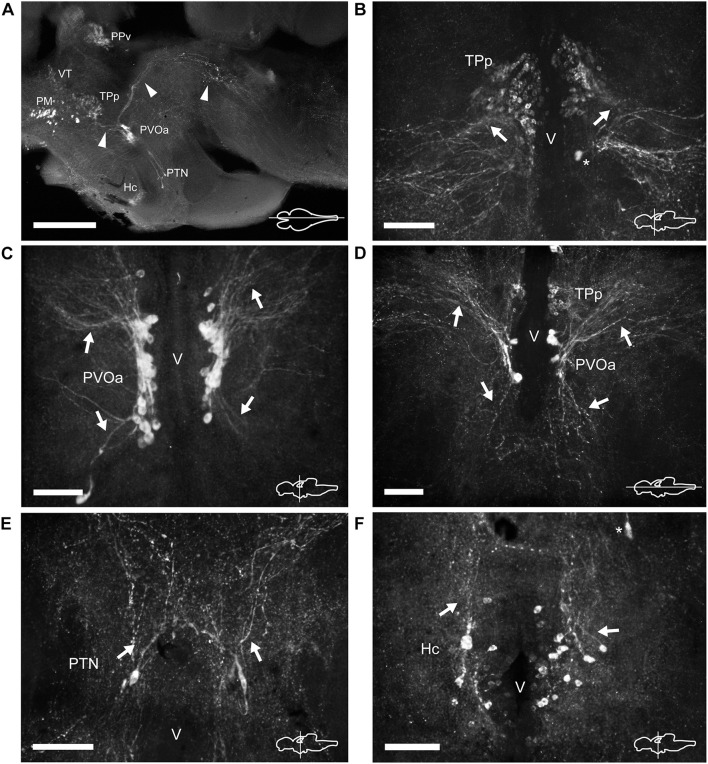
FIGURE 6.
TH immunoreactive neurons and fibers in the diencephalon of Nothobranchius furzeri at the level of the posterior tuberculum and hypothalamus. (A) Para-sagittal section showing a panoramic view of the distribution of all TH+ groups in the diencephalon. Arrowheads indicate TH+ fibers traveling from the paraventricular organ-accompanying cells toward the telencephalon and hypothalamus (bottom left), and across the mesencephalon to turn posteriorly toward the medulla (top right). (B) Transverse section showing the TH+ group in the periventricular nucleus of the posterior tuberculum. Arrows show the lateral tuft of projections emerging from the nucleus. The asterisk indicates a soma of the paraventricular organ-accompanying cells. (C) Transverse section showing the large pear shape neurons of the TH+ paraventricular organ-accompanying cells group. Arrows indicate the ascending and descending processes. (D) Horizontal section showing the relative position of the periventricular nucleus of the posterior tuberculum and the paraventricular organ-accompanying cells. Arrows point to ascending/descending processes of the paraventricular organ-accompanying cells. (E) Transverse section showing TH+ neurons at the posterior tuberal nucleus. The few cells of this group are positioned at each side of the ventricle and send projections in the dorsal direction (arrows). (F) Transverse section showing the TH+ neurons at the caudal hypothalamus. The arrow indicates the orientation of processes. The asterisk indicates a cell of the posterior tuberal nucleus. For abbreviations see list. The orientation/level of sections is shown at the bottom right corner of each panel. Dorsal is to the top in transverse sections. Rostral is to the top and left, in horizontal and para-sagittal sections, respectively. Scale bars, 500 μm (A) and 100 μm (B–F).

