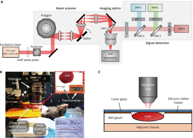
Fig. 1. Intravital microscopy setup to image hepatic lipid droplets in the liver of live mouse. (A) Schematic of custom-built laser scanning 2-photon microscopy system. (B) Photograph of intravital liver imaging setup. Temperature of mouse body and heating pad was continuously monitored. Silicon rubber heater was use for the temperature maintenance of the liver under cover glass. (C) Schematic of liver placement during the intravital liver imaging.
BPF, bandpass filter; DBS, dichroic beam splitter; PMT, photomultiplier tube; SPF, Shortpass filter.

