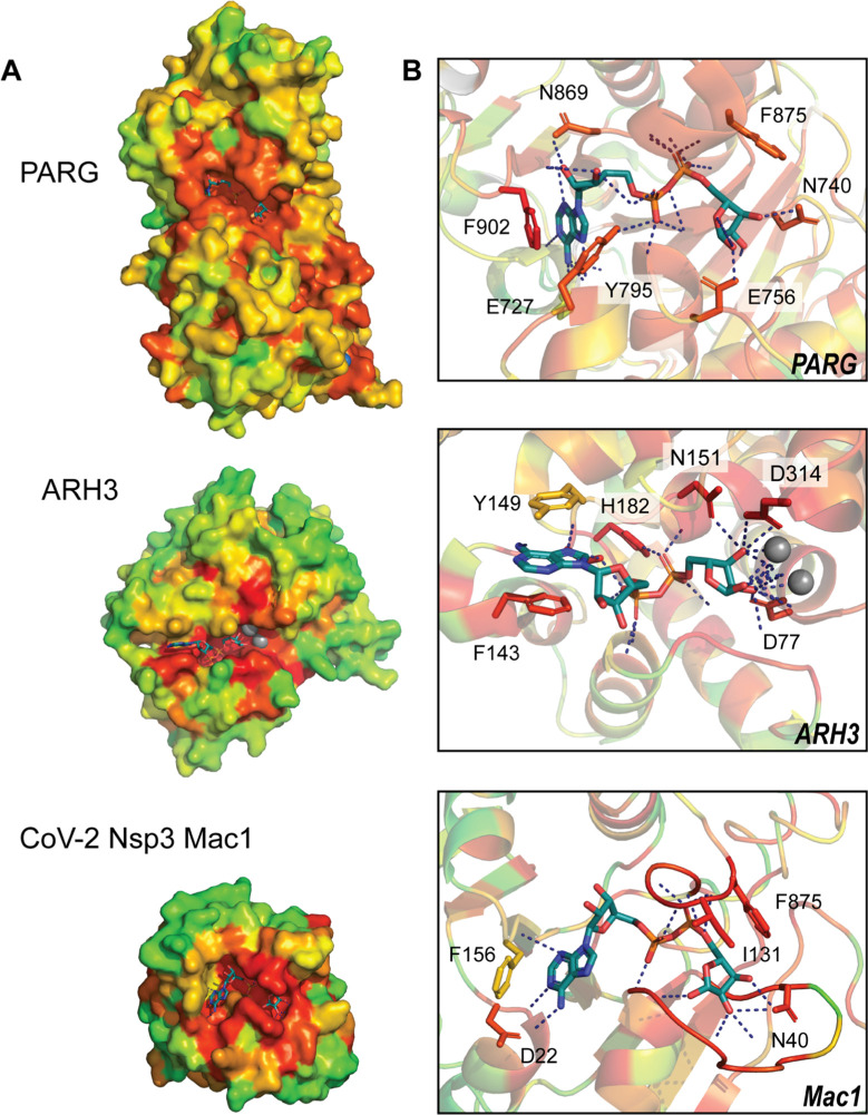
Fig. 2.
Evolutionary trace analysis identifies functional residues within PARG, ARH3, and CoV-2 Nsp3 Mac1 active sites. High-ranking ET values (red-orange) cluster at the active sites of each enzyme. Inspection of residues within the active sites (right panel) shows greatest conservation among residues coordinating the ADPr pyrophosphate linker and terminal ribose and increased variation within the adenine pocket.

