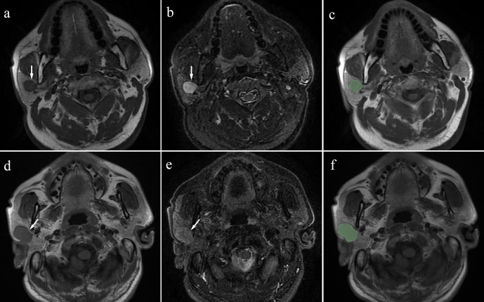
Figure 1.
a: T1-weighted image; b: fat-saturated T2 weighted image) Case 1: Pleomorphic adenoma in a 63-year-old male. A mass can be seen in the right parotid (arrow). (c) Manual segmentation of the mass. (d, e) (d: T1-weighted image; e: fat-saturated T2 weighted image) Case 2: Warthin’s tumour in a 51-year-old male. A mass can be seen in the right parotid (arrow). (f) Manual segmentation of the mass.

