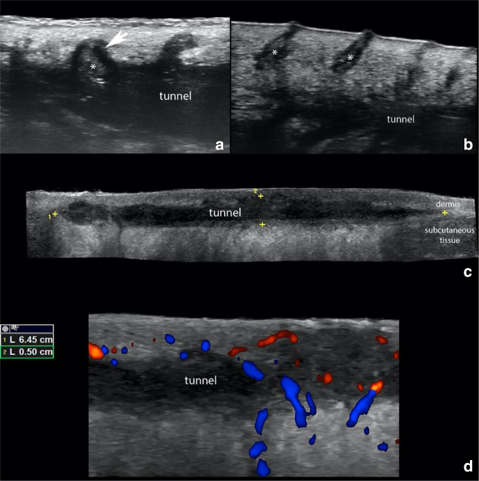
Figure 2.
Hidradenitis Suppurativa. (a) shows ballooning of a hair follicle (*, arrow) that is protruding into the periphery of a tunnel (donor of keratin sign). (b) presents dilation of the regional hair follicles (*). (c) demonstrate a 6.45 cm (length) x 0.5 cm (thickness) hypoechoic band-like structure that corresponds to a tunnel that runs in the dermis and upper subcutaneous tissue. (d) Doppler ultrasounds demonstrate hypervascularity in the periphery of the tunnel.

