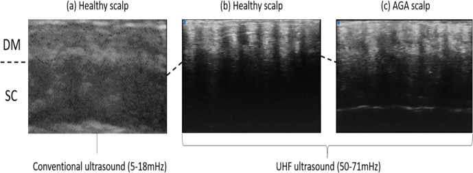
Figure 3.
The ultrasonographic images of conventional and newly developing ultrasound. (a) Healthy scalp depicted by conventional ultrasound. (b) Healthy scalp depicted by ultra-high-frequency (UHF) ultrasound. (c) Scalp of androgenetic alopecia depicted by UHF ultrasound. AGA, androgenetic alopecia; DM, dermis; SC, subcutis.

