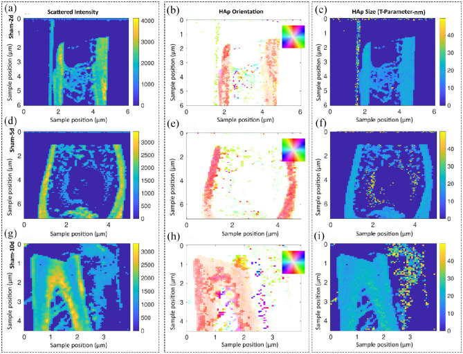
Figure 7.
Studying the collagen/hydroxyapatite (HAp) orientation and the size of hydroxyapatite plates in the sham group over time using small angle X-ray scattering (SAXS) analysis. Samples are scanned in a region of approximately 5 mm by 5 mm and each pixel corresponds to a probed region of 60 µm in radius. The shown information are a result of evaluating the scattering data and, therefore, test specific features at other length scales. Representative images of descriptively analyzing the scattered intensity (a, d, g) as well as the HAp orientation in degree (b, e, h) and size of the platelets in nm (c, f, i). No residuals of the defect were detected after day 10 for sham group (g, h). The HAp size was homogenous along the bone and around 2 nm for all time points (c, f, i). The color code in the HAp orientation analysis (b, e, h) shows the orientation degree, which corresponds to the inset, for example, red is along the y-axis of the image.

