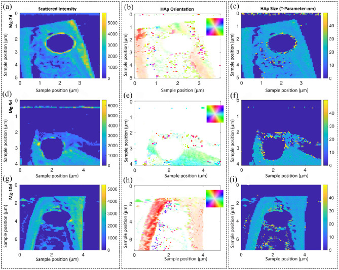
Figure 8.
Studying the collagen/hydroxyapatite (HAp) orientation and the size of hydroxyapatite platelets in the Mg-based alloy group over time using small angle X-ray scattering (SAXS) analysis. Samples are scanned in a region of approximately 5 mm by 5 mm and each pixel corresponds to a probed region of 60 µm in radius. The shown information are a result of evaluating the scattering data and, therefore, test specific features at other length scales. Representative images of descriptively analyzing the scattered intensity (a, d, g) as well as the HAp orientation in degree (b, e, h) and size of the platelets in nm (c, f, i). Only a minor degree of HAp orientation was observed at the Mg interface after 5 days (d–f). However, the HAp platelets size increased at the interface. The bone orientation at day 5 was in the horizontal direction; however bone was always orientated vertically at other time points. The color code in the HAp orientation analysis (a, d, g) shows the orientation degree, which corresponds to the inset, for example, red is along the y-axis of the image.

