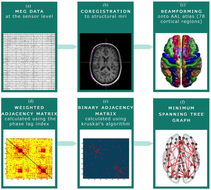
Figure 1.
MEG pre-processing steps. (a) MEG recording at sensor level. (b) The MEG recording was co-registered to the participants’ structural MRI. (c) Beamforming was applied to convert the MEG signal to source space: signals were projected onto the Automated Anatomical Labeling (AAL) atlas. (d) The phase lag index (PLI) was calculated between each of the 78 cortical regions of the AAL atlas. (e) The Minimum Spanning Tree (MST) was constructed based on the PLI, which consists of the 78 strongest connections. These connections were subsequently binarized. (f) An example of an MST graph.

