Fig. 4.
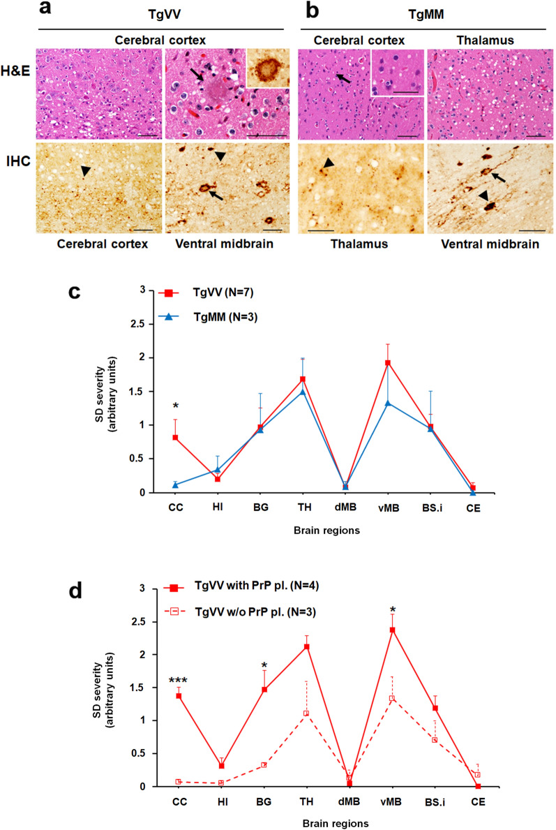
Hematoxylin and Eosin (H&E) staining and PrP immunohistochemistry (IHC). a, H&E (upper panels) & IHC (lower panels) staining of brain sections of TgVV mice inoculated with sPMCA-generated CWD-derived human PrPSc (Cd-HuPrPSc). Upper left panel: Spongiform degeneration (SD) preferentially affecting the deep layers of the cerebral cortex (CC). Upper right panel: Cortical eosinophilic plaque surrounded by vacuoles; inset: PrP IHC of the plaque shown in the upper right panel; the rim of the plaque was more heavily stained than its core. Lower left panel: PrP granules (arrowhead) accumulating mainly in the deep cortical layers. Lower right panel: Granular PrP inside and around the perikarya and processes (arrow) and plaque-like PrP deposits (arrowhead). b, H&E (upper panels) & IHC (lower panels) staining of brain sections of TgMM mice inoculated with sPMCA-derived human CWD prions. Upper left panel: SD (arrow) affecting less severely CC than thalamus (Upper right panel); inset in Upper left panel: higher magnification of SD. Lower left panel: Granular PrP deposits affecting the thalamus; Lower right panel: Pattern of PrP deposition similar to those shown in TgVV mice (Lower left panel in a) affecting the ventral midbrain. Bar size: 50 µm; antibody: 3F4. c, Profiles of brain distribution and severity of SD in the two Tg mouse lines challenged with Cd-HuPrPres were virtually identical, except for more severe lesions in the CC of TgVV mice. d, More severe lesions correlated with the presence of PrP plaques in TgVV mice; *P < 0.05–0.03, ***P < 0.006. HI: hippocampus, BG: basal ganglia, TH: thalamus, dMB and vMB: dorsal (d) and ventral (v) midbrain (MB), BS.i.: brainstem, inferior, CE: cerebellum. PrP pl.: PrP plaques; w/o: without. Each point of the lesion profile was expressed as mean ± standard error of the mean. Statistical significance was determined by a two-tailed Student’s t-test

