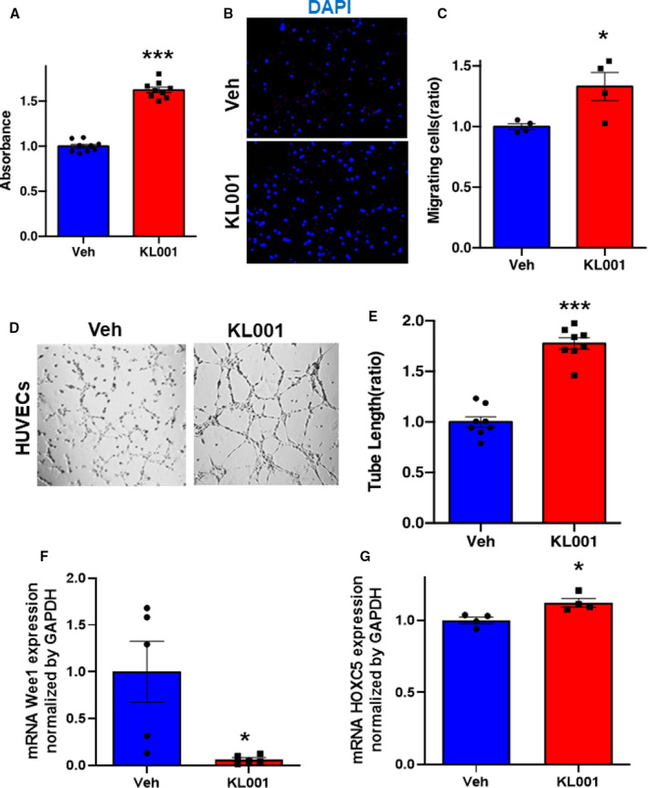
Figure 6. Stabilization of cryptochrome (Cry) 1 and Cry2 promotes angiogenesis in human umbilical vein endothelial cells (HUVECs).

A, WST‐1 proliferation assay of HUVECs in the response to the treatment with vehicle (Veh) or KL001 (40 μmol/L). Data are mean±SEM (n=8). ***P<0.001 vs Veh, analyzed by unpaired Student t test. B and C, Representative images of migrating cells stained by 4′,6‐diamidino‐2‐phenylindole (DAPI) and its quantitative analysis. Data are mean±SEM (n=4). *P<0.05 vs Veh, analyzed by unpaired Student t test. Bar=300 μm. Representative images of tube formation of HUVECs (D) and quantitative analysis by tube length (E) in each group. Data are mean±SEM. ***P<0.001 vs Veh, analyzed by unpaired Student t test. Bar= 1 mm. F and G, The expressions of mRNA Wee1 and HOXC5 in HUVECs treated with vehicle or KL001 (40 μmol/L) by quantitative polymerase chain reaction. Data are mean±SEM. *P<0.05 vs Veh, by unpaired Student t test.
