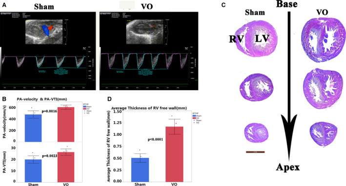
Figure 2. Volume overload (VO) increased in the abdominal aorta and inferior vena cava fistula model.

A, The representative echo image showed that 2 weeks after puncture, the pulmonary artery velocity and velocity time integral in the VO group increased. B, Quantitative statistics of pulmonary artery‐velocity and velocity time integral in sham and VO group, n=6, Student t‐test. C, Hematoxylin and eosin staining showed that 12 months after of puncture, the free wall of the right ventricle was thickened in the VO group, and the ventricular septum shifted to the left. D, Quantification of the thickness of the free wall of the right ventricle, n=6, Student t‐test. LV indicates left ventricle; PA, pulmonary artery; RV, right ventricle; VO, volume overload; and VTI, velocity time integral.
