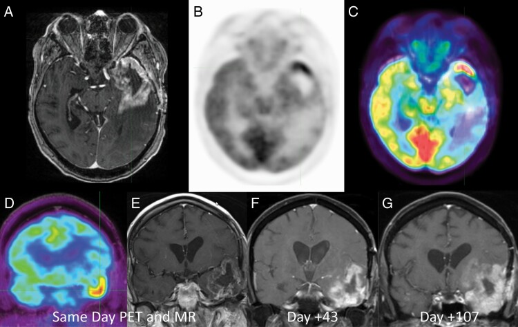
Figure 1.
44-year-old man with suspected recurrent glioblastoma after surgical resection and chemoradiation therapy 8 months prior. Axial post-contrast T1-weighted MR (a), axial 2-deoxy-2[18F]fluoro-d-glucose positron emission tomography (FDG PET) (b), and axial fused FDG PET-MR (c) images demonstrated intense FDG uptake along the temporal pole greater than adjacent cortex indicating metabolically active recurrent disease. Patient was deemed an unsuitable surgical candidate and followed with serial MRI examination. Coronal FDG PET (d) and coronal post-contrast T1-weighted MR (e) and subsequent short-term follow-up MRIs (f, g) demonstrate rapid progression of the disease.

