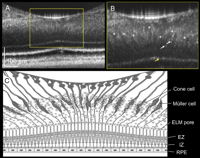
Figure 4.
Foveal Müller cells elucidated by adaptive optics (AO)-optical coherence tomography (OCT). (A, B) The AO-OCT image and magnified image of the human fovea B obtained from a 60-year-old man. The AO-OCT system delineates funnel-shaped bodies running diagonally from the inner limiting membrane to the external limiting membrane (ELM; asterisk). (C) Illustration of the AO–OCT image of the fovea. The illustration shows the characteristic features of the foveal Müller cells in addition to the hyper-reflective dots in the outer nuclear layer B (white arrow) and pores in the ELM B (yellow arrow). ELM, external limiting membrane; EZ, ellipsoid zone; IZ, interdigitation zone; RPE, retinal pigment epithelium.

