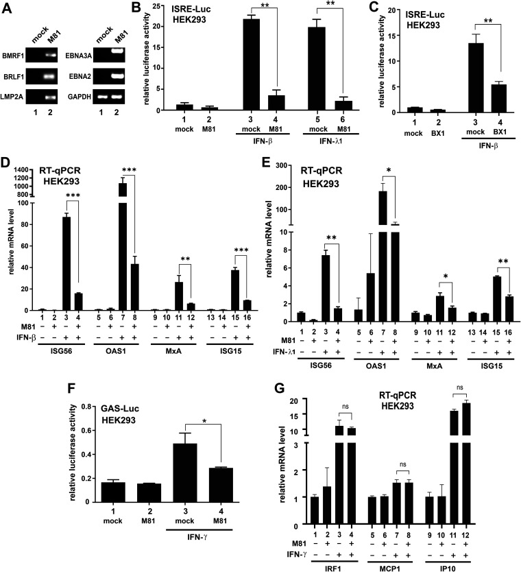
FIG 1.
Suppression of IFN signaling by EBV. The M81 or BX1 strain of EBV was used to infect HEK293 cells. Cells were treated with IFN after 24 h of infection and harvested after another 24 h. Error bars represent the SD (n = 3). (A) RT-PCR analysis of selected lytic and latent genes in M81-infected HEK293 cells. Cells were harvested 48 h after infection. (B and C) ISRE-Luc luciferase reporter assays. pISRE-Luc firefly luciferase reporter was transfected into HEK293 cells before infection, along with pSV-Rluc Renilla luciferase plasmid as an internal control. The ISRE promoter was induced with treatment of 1,000 U/ml IFN-β or 100 ng/ml IFN-λ1 for 24 h. Cells were lysed, and results are represented as the fold activation. The difference between mock- and M81- or BX1-infected samples was found to be statistically significant. (D and E) RT-qPCR analysis of ISG mRNAs responsive to IFN-β or IFN-λ1. Relative mRNA level was presented as the fold activation. The difference between mock- and EBV-infected samples was found to be statistically significant. (F) GAS-Luc reporter assay. Cells were treated with 50 ng/ml IFN-γ for 24 h. The results are represented as the fold activation. The difference between mock- and EBV-infected samples was found to be statistically significant. (G) RT-qPCR analysis of ISG mRNAs responsive to IFN-γ. The difference between mock- and EBV-infected samples was not significant (ns) statistically (P > 0.05).

