Figure 1:
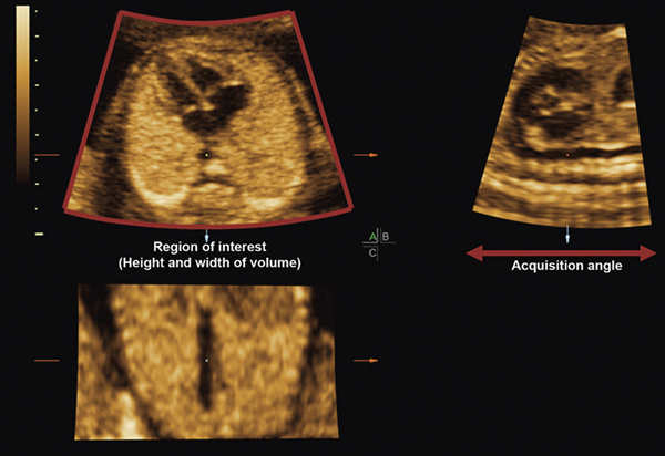
Multiplanar display of a STIC volume (normal fetal heart). The ROI box around the acquisition plane (apical four-chamber view) determines the height (y-plane) and width (x-plane) of the volume. Note that the box encompasses the entire fetal chest circumference. The B plane (sagittal image; upper right corner) demonstrates the acquisition angle of the volume, which determines the acquisition depth. Since the reference dot has been placed in the cross-section of the descending aorta in the A plane, the longitudinal descending aorta is visible in both the B and C planes.
ROI, region of interest; STIC, spatiotemporal image correlation.

