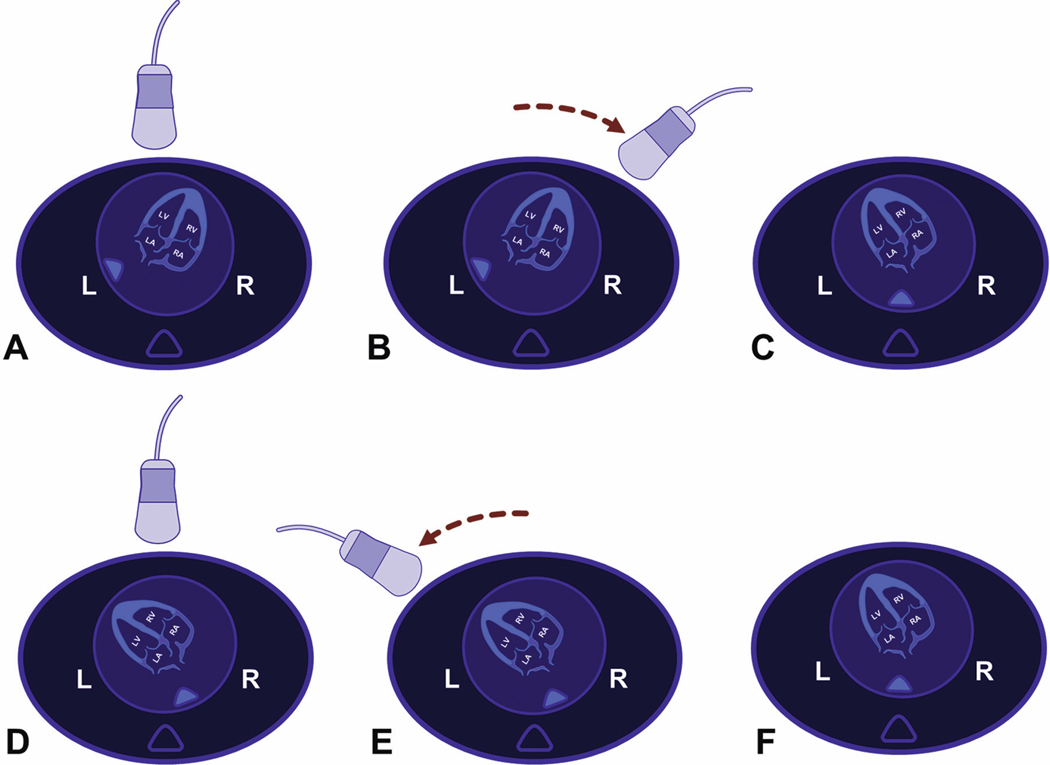
Figure 3:
“Driving the transducer” technique in a vertex fetus. A) Spine originally located at 8 o’clock in the apical four-chamber view; B) the transducer is driven on the maternal abdomen (toward the fetal right side) in a fixed arc until it lies above the cardiac apex; C) on the monitor screen, the fetal spine has “converted” to a more posterior position (6 o’clock), and the cardiac apex is now “up”; D) spine located at 5 o’clock in the apical four-chamber view; E) the transducer is driven leftward on the maternal abdomen (toward the fetal left side) in a fixed arc until it lies above the cardiac apex; F) on the monitor screen, the fetal spine has “converted” to a more posterior position (6 o’clock), and the cardiac apex is now “up.”
L, fetal left; R, fetal right.

