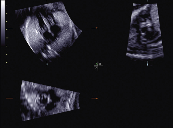
Figure 5:
STIC volume acquired from the subcostal four-chamber view, in which the spine was originally located at 4 o’clock. The acquisition plane (A plane) has been manually rotated on the z-axis so that the spine location is at 6 o’clock. As a result, the B plane image (ductal arch) becomes more blurred or “waxy” in appearance with diminished image clarity.
STIC, spatiotemporal image correlation.

