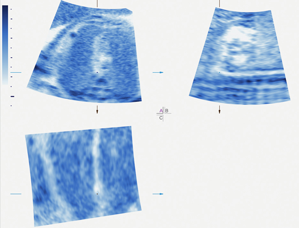
Figure 6:
During the STIC volume acquisition, fetal breathing occurred at the beginning, leading to a motion artifact in the area of the upper mediastinum. As a result, there is distortion of the 3-vessel and trachea view, as evidenced in the acquisition plane (upper left corner). Note that the pulmonary artery and ductus arteriosus (ductal arch view, B plane) appears distorted, and the anatomy cannot be assessed with confidence.
STIC, spatiotemporal image correlation.

