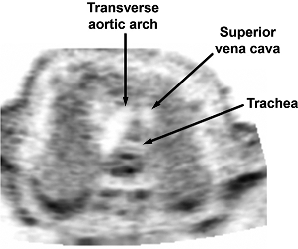
Figure 7:
Transverse view of the fetal upper mediastinum, demonstrating the transverse aortic arch (“dolphin”), cross-section of the superior vena cava, and cross-section of the trachea. Just immediately before beginning a STIC volume acquisition of the four-chamber view, one should tilt the transducer to ensure that the transverse aortic arch is also clearly visualized.
STIC, spatiotemporal image correlation.

