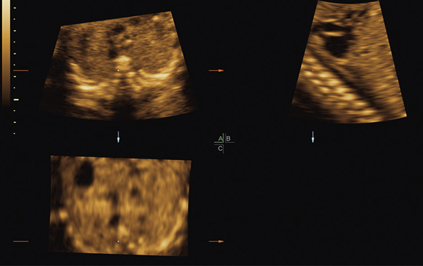
Figure 9:
Staircase spine (downstairs type). In the A plane (transverse view) of the STIC volume, note that the ossification centers of the spine appear “stacked” upon each other like a staircase or a caterpillar, and a coronal view of the curved ribs can be seen because the ossification centers are being imaged obliquely. The staircase spine is confirmed in the B plane, which shows the fetus inclined vertically downward. During a STIC volume acquisition, the ossification centers will appear to be “moving” in a vertical direction on the monitor screen.
STIC, spatiotemporal image correlation.

