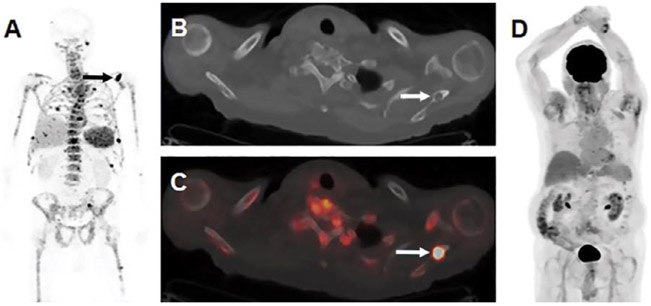
Figure 2.
Images show visualization of skeletal myeloma by using immuno-PET antibody composed of native daratumumab labeled with positron-emitting radionuclide zirconium 89 (89Zr) through chelator deferoxamine (DFO), or 89Zr-DFO-daratumumab, in an 80-year-old man with osseous myeloma. (A) Maximum intensity projection (MIP) image from 89Zr-DFO-daratumumab PET/CT demonstrates multiple foci of osseous avidity, including left scapular focus (arrow). (B) Axial CT and, (C) fused PET/CT images from 89Zr-DFO-daratumumab PET/CT demonstrate left scapular focus localizes to lytic osseous lesion at CT (arrows). (D) MIP image from fluorine 18 fluorodeoxyglucose PET/CT 1 week prior fails to identify lesions seen at 89Zr-DFO-daratumumab PET/CT. (reprinted from Radiology, 2020 (ref. 31).

