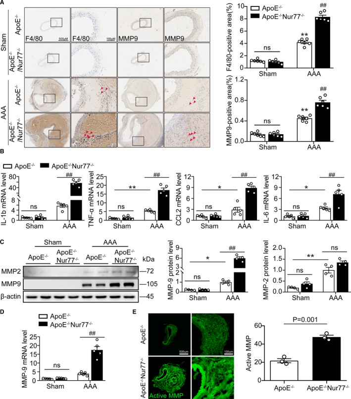
Figure 4. Nur77 deficiency enhances AAA lesion macrophage infiltration and inflammatory response.

A, Representative immunohistochemical staining images showing macrophages (F4/80) and MMP‐9 in mouse abdominal aortas, with the quantification results in the right panels (n=6 per group). B, The q‐PCR analysis of inflammatory cytokines (IL‐1b, TNF‐α, CCL2, and IL‐6) in the aortic wall (n=5 mice per group). C, Western blot analysis and quantitative results of MMP9 and MMP2 (n=4 mice per group). D, Gene expression of MMP9 in AAA lesioned tissues (n=4 mice per group). E, In situ zymography for gelatinase activity (n=3 per group). *P<0.05 vs Sham‐ApoE‐/‐ mice, **P<0.01 vs Sham‐ApoE‐/‐ mice; # P<0.05 vs AAA‐ApoE‐/‐ mice, ## P<0.01 vs AAA‐ApoE‐/‐ mice. Data are presented as mean ± SEM. Two‐way ANOVA followed by Tukey's multiple comparisons test for A–D. Student's two‐tailed t test for E. CCL2, chemokine (C‐C motif) ligand 2; IL‐1b, Interleukin‐1β; IL‐6, Interleukin‐6; MMP, matrix metalloproteinase; ns, nonsignificant; TNFα, tumor necrosis factor‐α.
