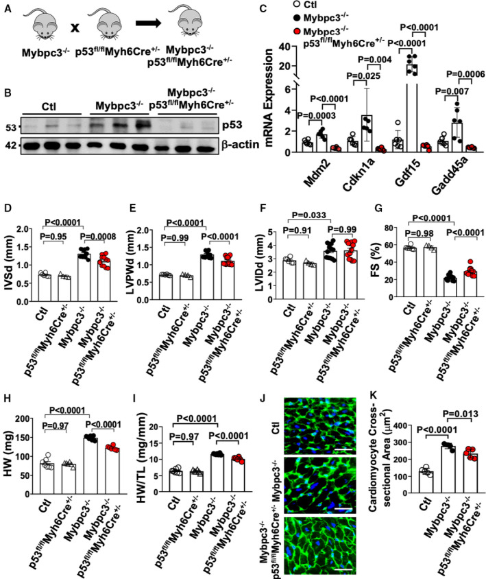
Figure 5. Deletion of cardiomyocyte p53 reduces pathological myocardial remodeling in Mybpc3−/− cardiomyopathy.

(A) Schematic of Mybpc3−/−/p53fl/flMyh6Cre+/− double‐null murine model generated by crossing a p53fl/flMyh6Cre+/− mouse with an Mybpc3−/− mouse. (B) Western blot of p53 from control (Ctl) (n=3), Mybpc3−/− (n=3), and Mybpc3−/−/p53fl/flMyh6Cre+/− (n=3) myocardial tissue at postnatal day (P) 25. Western blot of β‐actin used as loading control. (C) Measurement of p53 target gene expression (mouse double minute 2 [Mdm2], cyclin‐dependent kinase inhibitor 1 [Cdnk1a], growth differentiation factor 15 [Gdf15], and growth arrest and DNA damage inducible α [Gadd45a]) at P25 in Ctl (n=6), Mybpc3−/− (n=6), and Mybpc3−/−/p53fl/flMyh6Cre+/− (n=6) left ventricular (LV) tissue RNA. The genes of interest were normalized to Rpl32 expression. Fold changes are shown relative to Ctl gene expression. Echocardiography assessment of interventricular septal thickness at end diastole (IVSd) (D), LV posterior wall thickness at end diastole (LVPWd) (E), LV internal diameter at end diastole (LVIDd) (F), and fractional shortening (FS) (G) in Ctl (n=6), p53fl/flMyh6Cre+/− (n=4), Mybpc3−/− (n=13), and Mybpc3−/−/p53fl/flMyh6Cre+/− (n=13) mice at P25. Heart weight (HW) (H) and HW/tibia length (TL) ratio (I) from Ctl (n=7), p53fl/flMyh6Cre+/− (n=6), Mybpc3−/− (n=7), and Mybpc3−/−/p53fl/flMyh6Cre+/− (n=6) mice at P25. (J) Representative immunohistochemical staining using wheat‐germ agglutinin (green) and 4′,6‐diamidino‐2‐phenylindole (blue) of LV tissue from Ctl, Mybpc3−/−, and Mybpc3−/−/p53fl/flMyh6Cre+/− mice. Bar=10 µm. (K) LV cardiomyocyte cross‐sectional area from Ctl (n=5), Mybpc3−/− (n=5), and Mybpc3−/−/p53fl/flMyh6Cre+/− (n=5) mice. Minimum 50 cells/sample measured. All results are shown as mean±SEM.
