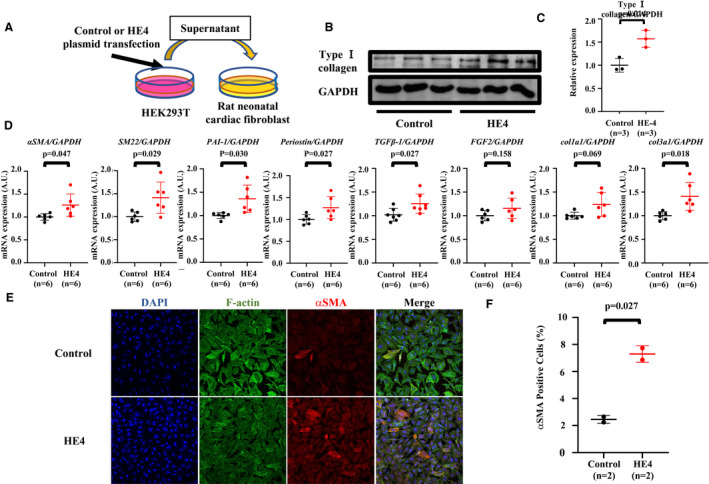
Figure 3. Cell‐secreted HE4 (human epididymis protein 4) induced fibroblast activation and extracellular matrix deposition in cardiac fibroblasts.

A, Experimental scheme for HE4 overexpression and transfer to cardiac fibroblast. B, Western blotting (WB) for type Ⅰ collagen in whole cell lysate of cardiac fibroblast stimulated by culture medium of human embryonic kidney 293T (HEK293T) cells. GAPDH was used as an internal control. C, Quantification of WB analysis. D, Quantitative reverse transcription–polymerase chain reaction analysis in cardiac fibroblast cultured with supernatant of HEK293T cells. The measurements were standardized to expression of the GAPDH. E, Overlay of images of cells stained for 4′,6‐diamidino‐2‐phenylindole (DAPI) (blue), F‐actin (green), and α‐smooth muscle actin (αSMA) (red). F, Quantification of αSMA‐positive cells as percentage of all cells. n=2 for control, n=2 for HE4. Unpaired t tests with Welch correction were used to compare groups. Col1a1 indicates collagen 1a1; col3a1, collagen 3a1; FGF2, fibroblast growth factor 2; PAI‐1, plasminogen activator inhibitor‐1; SM22, smooth muscle protein 22; and TGF‐β1, transforming growth factor‐β1.
