
Figure 4. HE4 (human epididymis protein 4) facilitated cardiac fibroblast activation through extracellular signal‐regulated kinase (ERK) signaling.
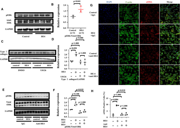
A, Western blotting (WB) for ERK in cardiac fibroblasts treated with human embryonic kidney 293T (HEK293T) culture medium. B, Quantification of phosphorylated ERK (pERK)/total ERK WB analysis. C, WB for type Ⅰ collagen in whole cell lysate of cardiac fibroblast stimulated by culture medium of HEK293T cells, and U0126 or dimethyl sulfoxide (DMSO). GAPDH was used as an internal control. D, Quantification of WB analysis. E, WB for ERK in whole cell lysate of cardiac fibroblast stimulated by culture medium of HEK293T cells, and anti‐HE4 antibody or IgG. GAPDH was used as an internal control. F, Quantification of WB analysis. G, Overlay of images of fibroblast cultured with supernatant of HEK293T cells, and anti‐HE4 antibody or IgG stained for 4′,6‐diamidino‐2‐phenylindole (DAPI) (blue), F‐actin (green), and α‐smooth muscle actin (αSMA) (red). H, Quantification of αSMA‐positive cells as percentage of all cells. n=3 for control, n=3 for HE4. Unpaired t tests with Welch correction and multiple comparison correction (Bonferroni method) were used to compare each group.