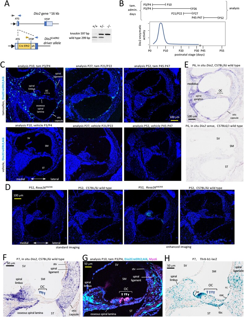
Figure 1.
Dio2 CreERt2 allele and its expression in the cochlea. A, The CreERt2 knockin displaces the first Dio2 coding exon. Triangle, promoter; dark blue. boxes, coding exons; light blue, untranslated regions; pA, poly(A) site after CreERt2 stop. Arrowheads denote genotyping primers; gel shows genotyping results. B, Examples of tamoxifen (tam) treatments relative to a reference diagram of cochlear Dio2 activity, modified with permission from our data in ref #16 (16); Copyright (2000) National Academy of Sciences, USA. C, Specific fluorescent cells (pale blue) in the otic capsule and in fibrocyte areas (spiral limbus, spiral ligament) in Dio2CreERt2;Ai6 mice at P10 after administration of tam at P3/P4 (mid-cochlear turn). Later administration at P21/P22 (mid-basal turn) and at P45, P46 and P47 (mid-turn) yielded very few Dio2+ cells. Corn oil vehicle or no treatment gives no specific signal at any stage tested. DAPI (dark blue), general nuclear stain. D, Lack of specific fluorescent signals in Rosa26Ai6/Ai6 or wild-type C57BL6/J control mice without treatment at P52, using standard confocal imaging (equivalent to panel C) (cochlear mid-turns). Enhanced imaging reveals weak background in the spiral limbus in Rosa26Ai6/Ai6 but not wild-type mice, suggesting low-level expression of Ai6 reporter in the absence of the Dio2CreERt2 allele. E, In situ hybridization analysis of Dio2 RNA location using antisense (top) and sense control (bottom) probes, in wild-type mice at P6. F, Magnified view of Dio2 in situ hybridization signals at P7 showing correlation with Dio2+ fluorescent cells in panel G. G, Magnified view of Dio2+ cells in the spiral ligament, spiral limbus, the otic capsule and osseous spiral lamina. No Dio2+ cells were detected in the organ of Corti (OC) and adjacent epithelia. Myo6 labels hair cells (arrowheads). H, Thrb receptor gene expression in organ of Corti and adjacent tissues that lack Dio2 expression. Abbreviations: ise, inner sulcus epithelium; OC, organ of Corti; ose, outer sulcus epithelium; rc, root cell area; SM, scala media; ST, scala tympani; stv, stria vascularis; SV, scala vestibuli; tbc, tympanic border cells.

