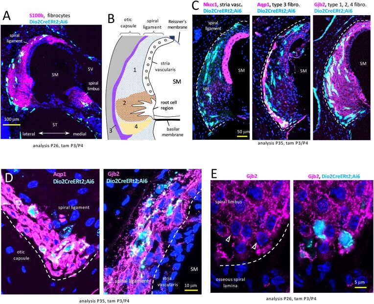
Figure 2.
Dio2+ fibrocytes in the spiral ligament (lateral) and spiral limbus (medial) in Dio2CreERt2;Ai6 mice. A, Fibrocyte regions indicated by S100b stain; cochlear mid-turn. B, Diagram of zones of fibrocyte types 1–4 in the spiral ligament. C, Lateral wall sections reveal that Dio2+ cells (pale blue) are not in the stria vascularis (NKCC1) but stain with markers of type 3 (Aqp1) and other fibrocyte types (Gjb2) (magenta). Double-positive cells appear whitish-blue. D, Magnified views of Dio2+ fibrocytes stained for Aqp1 (type 3, lower region) and Gjb2 (type 1, near stria vascularis). E, Spiral limbus Dio2+ fibrocytes stained for Gjb2 (arrowheads). DAPI (dark blue), general nuclear stain in panels A, B, D, E. Abbreviations: SM, scala media; ST, scala tympani; SV, scala vestibuli.

