Figure 7.
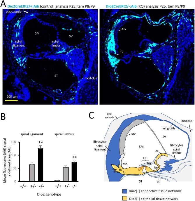
Response of Dio2+ cell types to Dio2-deficiency. A, Elevated Ai6 reporter expression in Dio2 knockouts (Dio2CreERt2/-) compared with control (Dio2CreERt2/+) mice. Signals increased in spiral ligament and spiral limbus fibrocytes, osteoblasts, and cells around the otic capsule and modiolus surfaces. Sections of mid-basal cochlear turns. B, Ai6 (Zsgreen1) fluorescent signal in fibrocyte areas in Dio2 knockouts (n = 3 mice; 9 or 10 views/group). Heterozygous and homozygous genotypes carry an equivalent, single Dio2CreERt2 allele. Pairwise comparison of -/- and +/+ groups, ** P < 0.001, Student t test; (+/+ control shows minimal background signal). C, Model for fibrocyte-mediated, paracrine-like control of T3 signaling. Dio2+ cells within the “connective tissue gap junctional network” (blue) may amplify T3 to transfer across the cellular boundary to the “epithelial tissue gap junctional network” (yellow) including T3-sensitive target cells in the vicinity of the organ of Corti. Hair cells (red outline) lack gap junctions with surrounding epithelial tissue. Dio2+ osteoblasts are not indicated in this simplified diagram. Abbreviations: idc, interdental cells; ise, inner sulcus epithelium; OC, organ of Corti; ose, outer sulcus epithelium; osl, osseous spiral lamina; rc, root cells; SM, scala media; ST, scala tympani; stv, stria vascularis; SV, scala vestibuli; tm, tectorial membrane.

