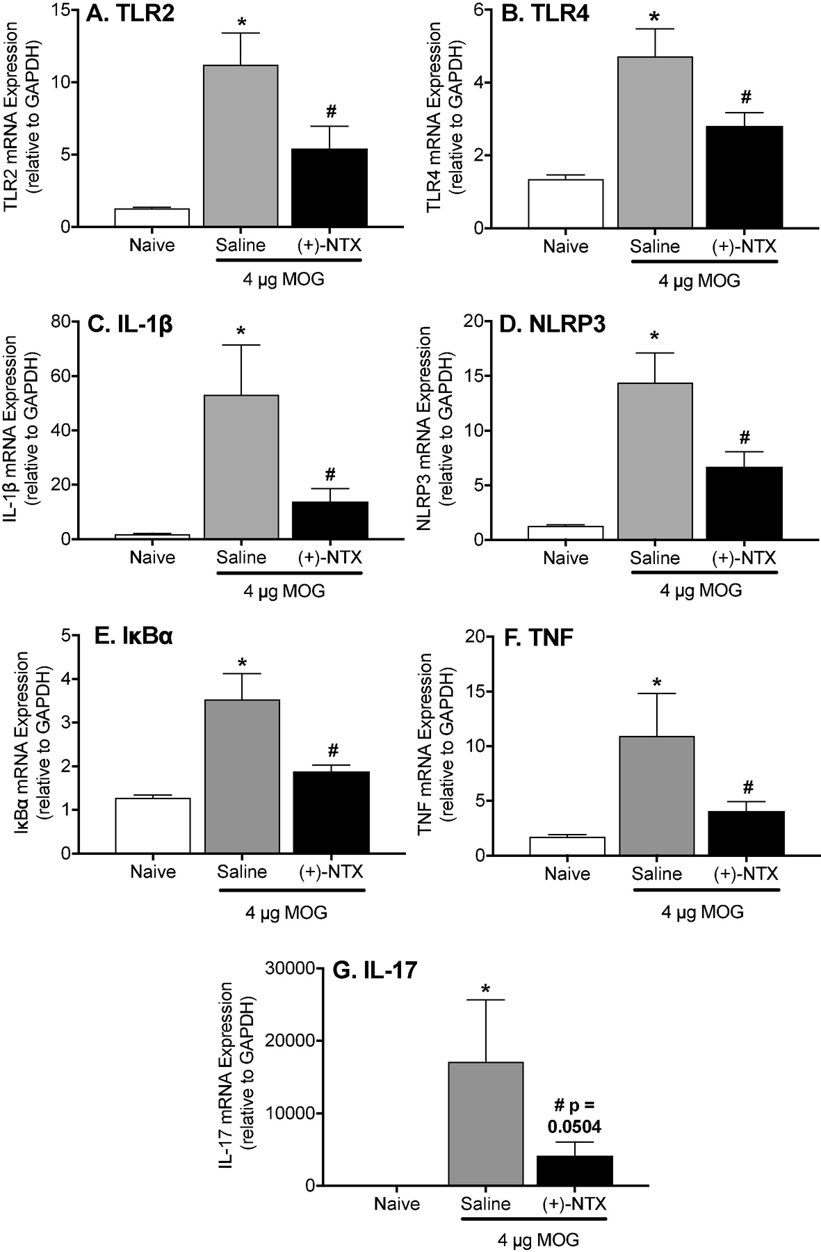
Figure 7. Repeated systemic administration of the TLR2/TLR4 antagonist (+)-Naltrexone [(+)-NTX], beginning 16 days after intradermal MOG, with tissue collection after 10 days of (+)-NTX treatment, suppresses EAE-induced neuroinflammatory markers in dorsal spinal cord of male Dark Agouti rats.

Male Dark Agouti rats received intra-dermal low-dose (4 µg) myelin oligodendrocyte glycoprotein (MOG). (+)-NTX (6 mg/kg subcutaneously [SC] 3x/day) vs. saline was initiated on Day 16 post-MOG, continuing for 10 days at which time tissues were collected for analysis. Each proinflammatory product (A: TLR2; B: TLR4; C: IL-1β; D: NLRP3; E: IκBα (reflective of NFκB activation); F: TNF-α; G: IL-17) was increased in response to EAE (1-way ANOVA comparing naïve to MOG+saline; IL-1β: p<0.005; NLRP3: p<0.001; TLR4: p<0.0001; TLR2: p<0.0005; IL-17: p<0.01; IκBα p<0.0001; TNF-α: p<0.005), and suppressed by (+)-NTX (1-way ANOVA comparing MOG+saline to MOG+(+)-NTX; IL-1β: p<0.05; NLRP3: p<0.05; TLR4: p<0.05; TLR2: p<0.05; IL-17: p=0.05; IκBα: p<0.001; TNF-α: p<0.05).
