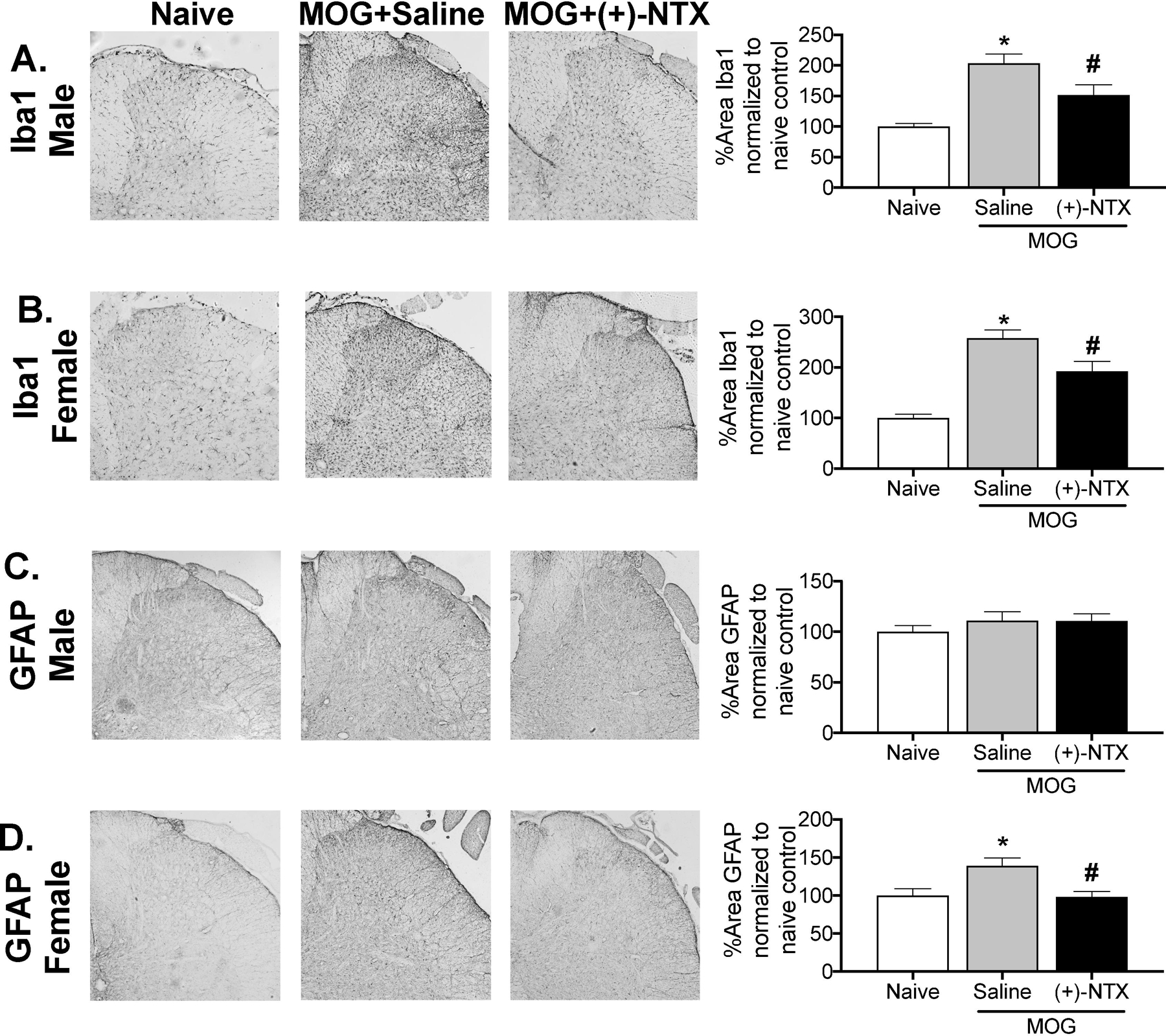
Figure 8. MOG increases the expression of glial immunoreactivity markers in male and female Dark Agouti rats, effects suppressed by (+)-NTX.

Spinal cord tissues from male and female Dark Agouti rats of Experiments 1 and 2 were collected for analysis here, upon completion of behavioral testing. For male Dark Agouti rats, MOG increased expression of the microglial immunoreactivity marker (Iba1) relative to naives in the dorsal horn of the lumbar spinal cord (Panel A; 1-way ANOVA comparing naïve to MOG+saline: p< 0.0001). This MOG-induced enhancement of Iba1 expression in males was reduced by (+)-NTX (1-way ANOVA comparing MOG+saline to MOG+(+)-NTX: p< 0.05). There was no increase in astrocyte GFAP expression in the dorsal horn of the lumbar spinal cord in males in response to MOG, nor was expression suppressed by (+)-NTX (Panel C). For female Dark Agouti rats, MOG again increased expression of the microglial immunoreactivity marker (Iba1) relative to naives in the dorsal horn of the lumbar spinal cord (Panel B; 1-way ANOVA comparing naïve to MOG+saline: p< 0.0001). As for males, this MOG-induced enhancement of Iba1 expression in females was reduced by (+)-NTX (1-way ANOVA comparing MOG+saline to MOG+(+)-NTX: p< 0.001). Unlike males, MOG increased expression of the astrocyte immunoreactivity marker GFAP relative to naives in the dorsal horn of the lumbar spinal cord (Panel D: 1-way ANOVA comparing naïve to MOG+saline: p< 0.05), an effect again reduced by (+)-NTX (1-way ANOVA comparing MOG+saline to MOG+(+)-NTX: p<0.01). For both male and female rats, n=3–9/group with 2–4 pictures analyzed/subject.
