
FIGURE 1.
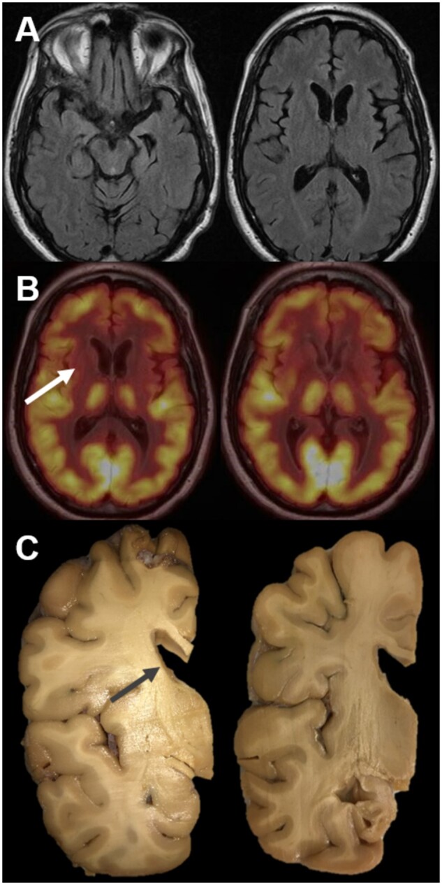
Neuroimaging and gross findings. Magnetic resonance imaging (A). Representative axial T2 Fluid Attenuated Inversion Recovery images showing mild global atrophy and also mild left temporal and left frontal segmental atrophy in this 56-year-old man with a 4-year disease history. Fluorodeoxyglucose positron emission tomography (FDG-PET) (B). Representative images showing normal symmetric cortical FDG avidity but with markedly decreased FDG avidity in the caudate and putamen bilaterally with only minimal avidity retained in the left posterior putamen. Gross examination (C). Coronal sections revealed dilation of the lateral ventricles with severe neostriatal atrophy, most prominent in the caudate (arrow).