. 2021 Sep 1;10:e68578. doi: 10.7554/eLife.68578

Figure 7. The closure of VFT leading to the rearrangement of inter-7TMDs.

(A) Front view of CaSRfully inactive CRDs and 7TMDs (cyan). (B) Front view of CaSRagonist+PAM CRDs and 7TMDs (purple). (C) The alignment of the part of CRD and 7TMDs in both fully inactive and agonist+PAM bound CaSR. (D) The alignment of inactive and agonist+PAM bound 7TMDs from top view. (E–G) The 7TMDs interface in the fully inactive state of CaSR is mediated by TM5 and TM6 (cyan) from top view and that of the agonist+PAM state is driven by TM6 from top view. Superposition of 7TMD of the inactive (cyan) and agonist+PAM bound CaSR (purple) show the rotation of 7TMDs. (H–J) Dose-dependent intracellular Ca2+ mobilization expressing WT (black dots) and mutant (red dots) CaSR (Figure 7—source data 1). The single mutations of F789A (H), F792A (I), and P823R (J) were designed based on the inactive density map. For (H–J), N = 4, data represent mean ± SEM.

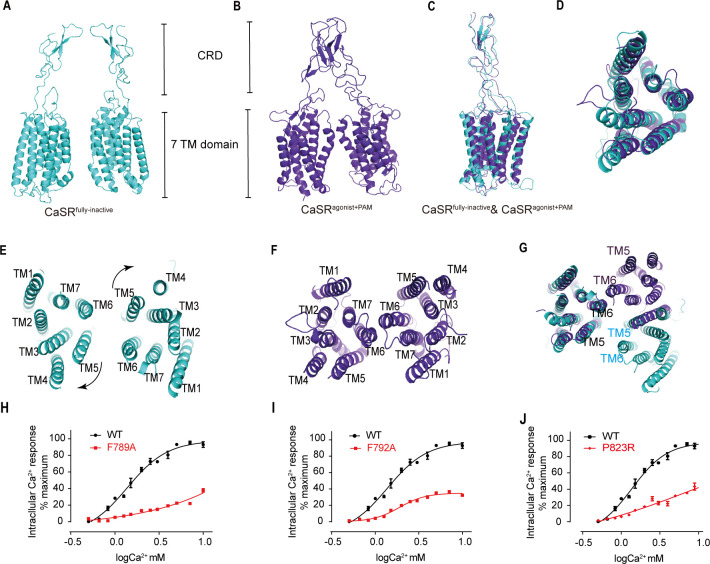
Figure 7—source data 1. Intracellular Ca2+ flux assay on various CaSR mutations.

Figure 7.

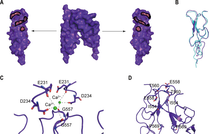
Figure 7—figure supplement 1. The homodimer interface of the CRDs in the active state of CaSR.

Figure 7—figure supplement 1.

(A) The homodimer interface of the CRDs (surface representation, purple). Contact regions (red) show residues within 4 Å of the opposite. It covers approximately 1079.09 Å2 of solvent accessible surface area. (B) There is a bound Ca2+ coordinated by carboxylate group of D234 and carbonyl oxygen of E231 and G557, holding the interface that is required to activate the receptor. (C) The CR–CR contacts were maintained through the cross-subunit hydrogen bonds between T560 and E558, and hydrophobic interaction of I554 and P569.
Figure 7—figure supplement 2. Conformational change of the 7TMDs interface during activation.

Figure 7—figure supplement 2.

(A) The 7TMD configuration in the inactive state from front view. There are pairwise symmetrical undefined maps that link the extracellular and intracellular part of TM5 and TM6 in the 7TM dimer interface, blocking the association of 7TMDs could regulate the function of CaSR. (B, C) The 7TMDs interface of the active state of mGluR5 (C, PDB:6N51) and GABAB (D, PDB:7C7Q) have the interface contact with TM6.